Imaging & After Your Examination
On-site CBCT imaging and MRI referrals for TMJ/TMD diagnosis in downtown Toronto — followed by a clear, collaborative treatment-plan review with Dr. Yolanda Cruz before any treatment begins.
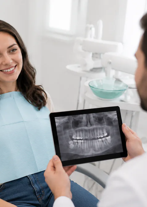
Based on the findings of your clinical TMJ examination, further diagnostic imaging may be recommended. Our office has on-site CBCT (cone beam computed tomography) imaging available, so in most cases this can be completed during your visit without travelling to a separate imaging centre. When soft-tissue imaging is required, Dr. Cruz will prepare an MRI requisition and refer you to an appropriate facility. After all findings are gathered, the diagnosis and treatment options are reviewed with you in plain language — collaboratively, and with time to ask questions before any treatment begins. We also accept referrals from other dental and medical providers for CBCT studies performed in our office. Schedule your evaluation or call 416-595-5490.
Imaging is matched to the clinical findings, not ordered by default
Diagnostic imaging is recommended only when the clinical examination raises a specific question that imaging can answer — for example, suspected hard-tissue changes in the condyle, possible disc displacement, airway anatomy, or asymmetric joint loading. It is not routine for every TMJ visit.
Two distinct modalities cover most TMJ/TMD diagnostic needs: CBCT for hard tissues (bone, teeth, condylar form, airway dimensions) and MRI for soft tissues (the articular disc, ligaments, joint effusion). Each images different structures, and the right choice depends entirely on what we need to see.
If you have prior imaging from another provider — a recent panoramic radiograph, a CBCT from your general dentist, or an MRI ordered by your physician — please bring it. Existing studies are reviewed first to avoid duplicate exposure.
Reviewed by Dr. Yolanda Cruz, DDS · Dr. Yolanda Cruz Dentistry On The Path · Toronto, ON
CBCT (cone beam computed tomography)
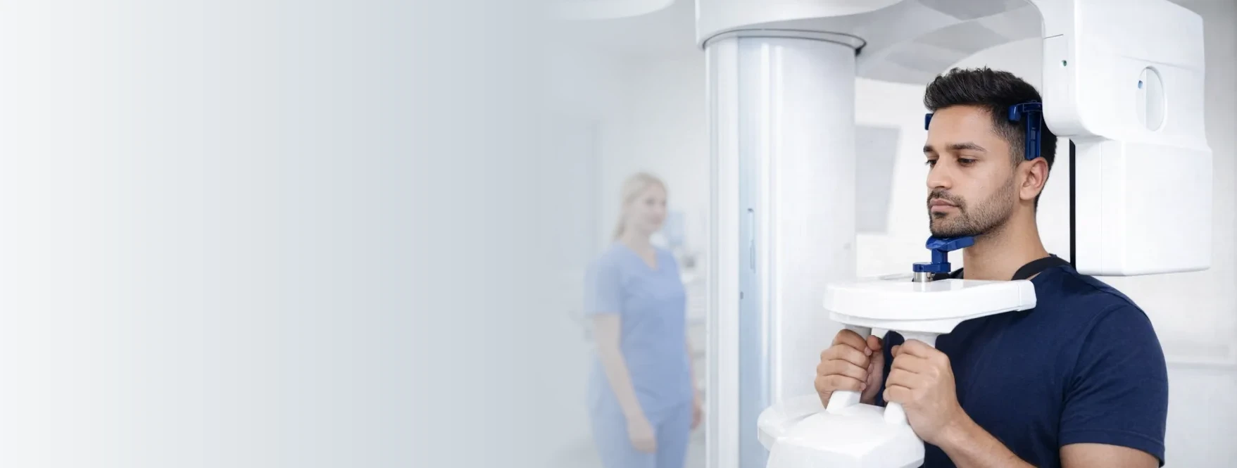
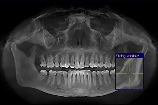
Our practice uses CBCT imaging for hard-tissue evaluation. CBCT is a compact version of a medical CT scanner that uses a cone-shaped X-ray beam to produce a three-dimensional image of the jaw joints, teeth, and upper airway. Scanning time is typically under 20 seconds, and the radiation dosage is substantially lower than a conventional medical CT scanner.
CBCT provides detailed images of hard tissues such as bone and teeth and is used to assess joint structure, condylar morphology, airway dimensions, and overall jaw anatomy. It does not image soft tissues such as the articular disc, ligaments, cartilage, or masticatory muscles — for those, an MRI is the appropriate study.
Because the CBCT is performed on-site, the scan, the immediate review, and the treatment-planning conversation often happen in a single visit. We also accept CBCT referrals from other dental and medical providers.
MRI for soft-tissue evaluation
When soft-tissue imaging is clinically indicated — most commonly to assess the condition and position of the TMJ disc, look for joint effusion, or evaluate the lateral pterygoid attachment — Dr. Cruz will prepare an MRI requisition and refer you to an imaging centre with TMJ MRI experience for the study.
A typical TMJ MRI includes both open- and closed-mouth sequences, which allow the disc to be assessed in both positions and any reduction (or non-reduction) of a displaced disc to be observed. MRI uses no ionizing radiation; the principal considerations are scan time (longer than CBCT) and any incompatible implants, which are screened for in advance.
Once the MRI report is returned, the images and the radiologist's findings are reviewed with you at a follow-up visit, and the working diagnosis is updated accordingly.
Imaging modalities compared
Not every patient needs imaging, and the right study depends on the clinical question. The table below is a general orientation only — the recommendation in your case is made by Dr. Cruz after the examination.
| Study | What it shows best | Ionizing radiation? | Typical indication in TMJ care |
|---|---|---|---|
| No imaging | Reserved for clinical-only cases | None | Mild, recent, muscle-dominant TMD where exam findings are clear |
| Panoramic radiograph (2D) | General overview of teeth, jaws, condyles | Low | Screening; often already available from a general dental visit |
| CBCT (3D, hard tissue) | Bone, condyle form, airway, dental anatomy | Low (well below medical CT) | Suspected condylar resorption, arthritic change, airway assessment, surgical-planning context |
| MRI (soft tissue) | Articular disc, ligaments, joint effusion | None | Suspected disc displacement (with or without reduction), persistent joint pain, before considering joint-directed procedures |
Reviewing findings & building your plan
Once the examination is complete — including any imaging — Dr. Cruz will review all findings with you clearly. You will understand what has been identified and what your treatment options are. All decisions are made collaboratively, and you will have time to ask questions and consider your options before any treatment begins.
- A walk-through of the imaging on screen, showing exactly what was found and what it means
- A working diagnosis explained in plain language — muscle-dominant, joint-dominant, bite-driven, or a combination
- A written treatment plan with the recommended sequence of steps
- A cost estimate for the plan, including any items typically covered by Canadian extended-health plans
- Time to take the plan home, ask follow-up questions, and consult others before committing
- A clear path for any referrals required (e.g., an oral and maxillofacial surgeon or a sleep physician) when indicated
Dr. Yolanda Cruz is a general dentist. All services provided at this practice are within the scope of general dentistry. When findings suggest the care of a specialist (oral and maxillofacial surgery, oral medicine, sleep medicine, ENT), Dr. Cruz coordinates the referral.
Is on-site imaging part of your visit?
Imaging or a post-exam review may be part of your TMJ appointment if any of the following apply:
- Persistent jaw pain or joint tenderness that hasn't resolved with conservative measures
- Clicking, popping, or locking — particularly when symptoms are recent or worsening
- Concern for arthritic change, condylar resorption, or asymmetric joint loading
- A history of jaw trauma, orthognathic surgery, or prior TMJ procedures
- Suspected disc displacement — with or without reduction — based on clinical findings
- An airway question (snoring, witnessed apneas, suspected sleep-disordered breathing) raised during exam
- Planning for joint-loaded restorative work or orthodontic treatment where TMJ status matters
- You were referred to our office by another dentist or physician for CBCT imaging
Imaging is generally not recommended when your symptoms are mild, recent, muscle-dominant, and the exam findings are clear-cut — in those cases a conservative trial is usually the appropriate first step, and imaging is reserved for non-response or progression.
What the imaging and review visit feels like
Most patients tell us they expected the imaging part to be more involved than it is. A typical CBCT and post-exam review involves:
- You stand or sit upright at the CBCT — no lying down, no contrast injection, no enclosed tunnel
- A lead apron is placed, your chin is positioned on a rest, and you bite gently on a guide
- The arm rotates around your head for roughly 15–20 seconds — you hear a mechanical sound and stay completely still
- Total scan time is under a minute including positioning; total room time is typically 5–7 minutes
- The images are reconstructed within a few minutes and reviewed with you on a large screen at the next chair
- Dr. Cruz walks through the findings, the working diagnosis, and the recommended next steps before you leave
No injections, no drilling, no appliance impressions on this visit unless the plan has already been agreed in advance. The review visit is diagnostic and educational — a written plan and cost estimate follow before any treatment begins.
What imaging adds to your treatment plan
Targeted imaging — used selectively and matched to the clinical question — sharpens the diagnosis and shapes a plan that is right for your joint, not a generic TMJ.
Same-visit answers
On-site CBCT means the scan, the read, and the diagnosis conversation can usually happen in a single appointment — no separate imaging-centre trip.
3D detail of the condyle
CBCT shows the condylar surface, joint space, and any bony changes in three dimensions — flat 2D panoramic films cannot.
Right modality for the right question
CBCT for bone, MRI for the disc — selecting (or sometimes combining) studies prevents both under-imaging and unnecessary exposure.
Low radiation dose
Modern dental CBCT delivers a small fraction of the dose of a conventional medical CT scan, with image quality suited to dental and joint anatomy.
Clear, collaborative plan
You see the same images Dr. Cruz sees. The diagnosis, options, costs, and any referrals are discussed openly before any treatment starts.
Open referral pathway
Other dentists and physicians can refer their patients to our office for CBCT — useful for implant planning, endodontic questions, or TMJ work-up.
Dr. Cruz's clinical note
"Imaging is something I order to answer a specific question — never to confirm something I already know clinically. If the exam tells me this is muscle-dominant TMD in a recent grinder, a CBCT isn't going to change my plan. If it tells me there's a possible condylar change or a disc problem, then we image — and we image the right tissue. CBCT for bone, MRI for the disc."
"What patients often find most useful isn't the scan itself — it's sitting down afterwards, looking at their joint on screen, and finally understanding what's been going on. That's the part I plan for. Imaging without a proper review conversation is half a service."
— Dr. Yolanda Cruz, DDS, Dr. Yolanda Cruz Dentistry On The Path, Downtown Toronto
Risks & considerations
- CBCT involves a low — but non-zero — dose of ionizing radiation; it is recommended only when the diagnostic benefit clearly outweighs the exposure
- CBCT does not image soft tissue (articular disc, muscles, ligaments) — a disc problem cannot be diagnosed on CBCT alone
- MRI does not image bone in detail — an arthritic or resorbing condyle is better assessed on CBCT
- MRI is contraindicated or requires special protocols for certain implanted devices; you will be screened beforehand
- Pregnant patients should inform the practice before imaging — non-urgent CBCT is typically deferred
- Imaging findings can be incidental and unrelated to the presenting complaint; we discuss these openly during the review
- A diagnosis is a working one — it can be refined as treatment response unfolds
Frequently asked questions about imaging & the post-examination visit
Imaging is recommended only when it will change the diagnosis or the plan. CBCT is used to evaluate hard tissues — the condyle, joint space, bony anatomy, airway — and MRI is used to assess soft tissues, primarily the articular disc. Mild, recent, muscle-dominant TMD usually does not require imaging; persistent, joint-dominant, or progressive cases often do.
A dental CBCT delivers a low dose of ionizing radiation — substantially lower than a conventional medical CT scan, and roughly in the range of a few panoramic dental radiographs depending on the field of view and settings selected. The scan takes under 20 seconds. Imaging is recommended only when the clinical question warrants it; pregnant patients should always inform the practice before any imaging.
In most cases yes. Because CBCT imaging is on-site, the scan and the immediate review can typically happen in the same visit. MRI cannot — it is performed at an external imaging centre after a requisition is prepared, and is reviewed at a follow-up visit once the report is returned.
Coverage varies. Many Canadian extended-health and dental plans cover a portion of dental diagnostic imaging including CBCT. MRI is typically arranged through the medical system and follows provincial healthcare rules in Ontario. We provide documentation for pre-authorization wherever possible, and the CDCP page outlines what is eligible under the federal plan.
Yes, in most cases. If the field of view captured the joints and the study is recent enough, prior CBCT data is reviewed first to avoid repeating imaging. Please bring the original DICOM data (a disc or secure link) — flat JPG screenshots are not usually sufficient for diagnostic review. The same applies to MRI studies done elsewhere.
The follow-up visit is a structured review. Dr. Cruz walks through the findings on screen, explains the working diagnosis in plain language, presents the recommended sequence of treatment, and provides a written cost estimate. Decisions are collaborative — you'll have time to ask questions, consider the plan, and consult others before any treatment is started. For practical location and access details see the parking & directions page.
Medical Disclaimer
This content is for informational purposes only and does not constitute dental or medical advice, diagnosis, or treatment. Imaging recommendations and post-examination plans depend on individual clinical findings and history. CBCT and MRI carry their own indications, contraindications, and considerations. Consult Dr. Yolanda Cruz or another qualified dental professional regarding your symptoms, imaging options, and treatment. Individual results may vary.
Ready to schedule your TMJ examination in downtown Toronto?
Book a focused evaluation with Dr. Cruz. If imaging is needed, on-site CBCT is usually available the same day — and you'll leave with a working diagnosis and a written plan reviewed in plain language.