Advanced diagnostic tools for accurate TMJ assessment in Toronto
Accurate TMJ/TMD diagnosis requires objective information — not guesswork. At Dentistry On The PATH, Dr. Yolanda Cruz uses a suite of advanced diagnostic technologies to evaluate jaw joint function, bite forces, and surrounding structures. Together they give a precise picture of what's causing your symptoms, so your treatment plan is built on real data — not assumptions.
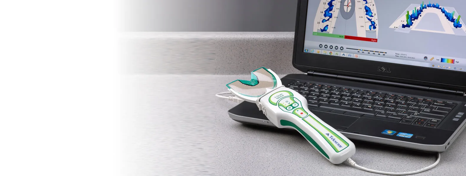
T-Scan is a digital system that records the timing and force distribution of your bite in real time.
This provides objective data about how the upper and lower teeth come together — information that can identify bite imbalances that may be contributing to your TMJ symptoms.
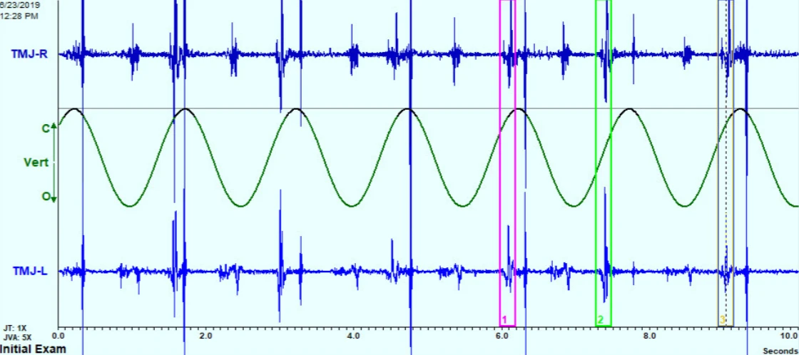
JVA is a non-invasive technology that records the vibrations produced by the jaw joint tissues during movement.
Different patterns of vibration correspond to different types of joint pathology — a healthy joint glides nearly silently, while a displaced disc, osteoarthritic joint, or perforated disc each generate distinct, frequency-specific signatures.
JVA can also be used to monitor changes in joint function over the course of treatment — making it useful for tracking progress as well as initial diagnosis.
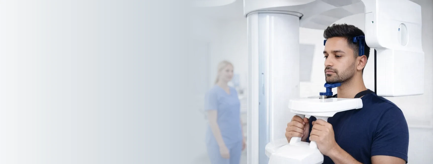
CBCT produces a three-dimensional image of the jaw joints, teeth, surrounding bone structures, and airway — all from a single scan.
The scanning time is typically under 20 seconds, with radiation dosage substantially lower than that of a medical CT scanner.
CBCT provides detailed imaging of hard tissues — bone and teeth. It does not image soft tissues such as cartilage or ligaments; for that, MRI is the appropriate next step.
When soft tissue imaging is clinically indicated — for example, to assess the condition of the TMJ disc — Dr. Cruz will prepare an MRI requisition and refer you to an appropriate imaging centre.
MRI complements CBCT by showing what the cone beam scan cannot: the articular disc, ligaments, muscles, and other soft tissues that play a critical role in jaw joint function.
Each technology captures a different dimension of your jaw joint function. Used together, they replace guesswork with data — so your treatment is built on what's actually happening, not what we assume.
T-Scan + JVA capture how your bite and jaw joint actually behave during movement — not just how they look at rest.
CBCT and MRI together cover both hard tissues (bone, teeth) and soft tissues (disc, cartilage, ligaments).
Data-driven diagnosis means treatment decisions are grounded in measurable findings — not pattern matching or guesswork.
Dr. Yolanda Cruz combines clinical expertise with advanced diagnostic technology to identify the root cause of your TMJ symptoms. Schedule a consultation at our Downtown Toronto office to begin a data-driven treatment plan.